Using AI to Achieve Earlier Diagnosis of Pancreatic Cancer
 Thought LeadersTakayuki BabaHuman Enhancement ProjectFujitsu Research
In this interview, we talk to Takayuki Baba from Fujitsu Research about ongoing research using artificial intelligence to achieve earlier diagnosis of pancreatic cancer.
Please could you introduce yourself and tell us about how you came to research artificial intelligence in the context of pancreatic cancer?
I am Takayuki Baba, and I am researching medical image diagnosis support technology as a “converging technology” that combines image analysis technology and medical science at Fujitsu Research. Converging technologies combine two or more different social sciences and technology areas to achieve a specific goal and represent a major focus of Fujitsu’s R & D.
Fujitsu Research has a track record in the research and development of technologies for the detection of multiple types of lesions on computed tomography (CT) images with AI and the retrieval of past CT images with a similar distribution of lesions, which are used in medical diagnostic imaging support technologies to help physicians make diagnoses.
Fujitsu and the Southern Tohoku General Hospital have started joint research with Fujitsu Japan Limited and FCOM CORPORATION on AI technology for detecting pancreatic cancer from non-contrast CT images through FCOM, which has been supporting the medical system of Southern Tohoku General Hospital.
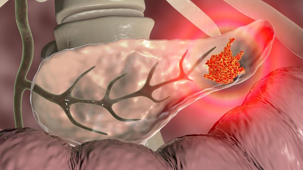
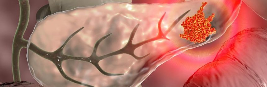
Image Credit: Kateryna Kon/Shutterstock.com
Why is the survival rate for pancreatic cancer lower than other cancers?
The survival rate for pancreatic cancer is said to be low, as it is often found when it has already progressed to a state that is difficult to treat.
Why is the early detection of pancreatic cancer a difficult task?
One of the reasons that makes the early detection of pancreatic cancer difficult is that there are few noticeable symptoms, and it is difficult to conduct voluntary examinations until the cancer progresses. Further, as the pancreas is at the deepest part of the body, a relatively simple imaging test, such as abdominal ultrasonography, cannot show the entire pancreas. It can be said that it is difficult to distinguish the area where cancer is suspected because the contrast between the lesion and the surrounding area is low in various image diagnoses such as CT, MRI, and PET.
What research has been carried out by Fujitsu and the Southern Tohoku General Hospital, and what is the goal of this research?
In April 2022, Fujitsu and Southern Tohoku General Hospital embarked on a joint research project to develop an AI technology that can detect areas of suspected pancreatic cancer from CT images without contrast agents (non-contrast CT scans) with the aim of detecting pancreatic cancer at an earlier stage.
This project aims to develop a technology for detecting suspected areas affected by pancreatic cancer by training an AI with approx. 300 anonymized CT scans of pancreatic cancer patients held by the Southern Tohoku General Hospital and offer an optimal image analysis method based on the shape of organs and cancer tumors.
With this technology, we anticipate that non-contrast CT scans, which have not been the subject of AI research until now but are widely used for medical checkups, can also be used to detect areas of suspected pancreatic cancer and enable doctors to refer patients to the gastroenterology department for detailed examination promptly. Our goal is to apply this technology to many CT scans worldwide.
How will this AI technology work and achieve earlier diagnoses of pancreatic cancer?
Pancreatic cancer is very difficult to detect. The low contrast of non-contrast CT scans makes it difficult to locate the pancreas itself, as boundaries between the pancreas and other organs are unclear. Fujitsu and Southern Tohoku General Hospital aim to develop an AI technology that can identify the region corresponding to the pancreas (part marked yellow in the image below) and detect the suspected parts affected by cancer (part marked red in the image below) by estimating the continuity between the anterior and posterior cross-sectional images in consideration of the anatomical tissue connection and automatically performs three-dimensional analysis including the anterior and posterior cross-sectional images in areas with strong continuity and planar analysis in areas with weak continuity.
In this way, slight visual discrepancies between a normal (healthy) pancreas and suspected parts affected by pancreatic cancer can be visualized on CT scans to detect patients with a high risk of pancreatic cancer. The technology thus aims to support doctors in providing more efficient and reliable visual diagnostic imaging and help them refer patients with a suspected risk of pancreatic cancer to the gastroenterology department for a detailed examination to detect pancreatic cancer at an early stage.
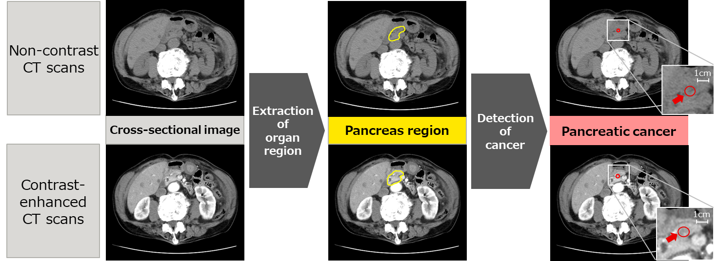
Figure: Detection of suspected pancreatic cancer using the newly developed technology
How will this technology improve the quality of life of pancreatic cancer patients?
By using this technology to support the early detection of pancreatic cancer, we expect to see a dramatic increase in the number of patients with pancreatic cancer who can be saved through treatment and surgery through early diagnosis of pancreatic cancer.
How could this research be applied across medical and health services to improve healthcare?
In the future, we will verify the effectiveness of the technology developed in this joint research and apply it to a wide range of clinical settings with the aim of saving lives and improving the quality of life of patients with pancreatic cancer.
What stage is this research currently at, and what is the next step?
Fujitsu and Southern Tohoku General Hospital are currently developing a technology to detect suspected parts affected by pancreatic cancer by training an AI with approx. 300 anonymized enhanced CT scans and non-enhanced CT scans held by the Southern Tohoku Hospital.
As a next step, we will apply this AI technology in clinical practice, first to detect tumors and pancreatic duct dilatation, which are typical findings of early pancreatic cancer but are difficult to detect, and then to test the effectiveness of the technology by detecting findings that require clinical follow-ups, such as cysts and local atrophy of the pancreas.
How do you believe artificial intelligence will influence medical research in the future, and what impacts will this have on healthcare?
At present, AI technology has made remarkable progress in the field of medical care, particularly in the area of diagnostic imaging support, and we believe that AI will be indispensable in various aspects of medical care in the future. In this joint research, we are conducting cutting-edge research on AI technology that detects pancreatic cancer from CT scans in order to detect pancreatic cancer at an early stage.
In the future, we believe that we can contribute to the creation of a society in which people can live healthily by widely applying the results of our research to medical care and health promotion services.

Image Credit: elenabsl/Shutterstock.com
Where can readers find more information?
Currently, readers can refer to our following press release: https://www.fujitsu.com/global/about/resources/news/press-releases/2022/0425-01.html
About Takayuki Baba
Takayuki Baba, Human Enhancement Project, Converging Technologies Laboratories, Research Unit, Fujitsu Research, Fujitsu Limited.
Current position: as a project manager at Fujitsu Research, I oversee the research and development group for cancer early detection technologies.
I joined Fujitsu in 1999 and have been engaged in research and development of machine learning, multimedia retrieval, and medical image diagnosis support technologies at Fujitsu Research. From 2009 to 2010, I was a Visiting Scientist at Cornell University in the United States where I studied image recognition technology using AI. From 2014 to 2018, I served as Visiting Associate Professor at the National Institute of Natural Sciences. In 2018, I became a part-time lecturer at Tokyo Institute of Technology. My specialty is medical image analysis (doctoral degree in engineering).
Posted in: Insights from Industry | Medical Science News | Medical Research News | Medical Condition News | Disease/Infection News | Healthcare News
Tags: Artificial Intelligence, Cancer, Computed Tomography, CT, Diagnostic, Gastroenterology, Healthcare, Hospital, Imaging, Machine Learning, Medical Research, Pancreas, Pancreatic Cancer, Research, Research Project, Surgery, Tomography
.png)
Written by
Emily Henderson
During her time at AZoNetwork, Emily has interviewed over 300 leading experts in all areas of science and healthcare including the World Health Organization and the United Nations. She loves being at the forefront of exciting new research and sharing science stories with thought leaders all over the world.
Source: Read Full Article