The Placenta, an Afterthought No Longer
The placenta may be dismissed as “afterbirth,” deemed an afterthought in discussions about pregnancy and even relegated, literally, to the trash bin. But at long last it is beginning to get its due.
In the past three weeks, scientists have published three significant studies of this ephemeral organ. One gave a detailed analysis of all the genes expressed, or converted into functioning proteins, in the placenta; another experimented with a way to silence that expression when it causes trouble. In the third, researchers created mini-placentas, three-dimensional clusters of cells, or organoids, that mimic the real thing in the lab, and can be used as models for studying it.
In addition, at a recent meeting in Baltimore of the Human Placenta Project, several teams of researchers showed off sophisticated new techniques that enable the placenta to be studied in real time. That work could help doctors diagnose dangerous complications in pregnancy — including pre-eclampsia (a form of high blood pressure), preterm birth and fetal growth restriction — early enough to intervene. It might also help to reveal why boys are much more vulnerable than girls to disorders of brain development, including schizophrenia, A.D.H.D., autism, dyslexia and Tourette syndrome.
“The missing link between complications during pregnancy and development of the fetal brain has been hiding in plain sight for a long time,” said Dr. Daniel R. Weinberger, director of the Lieber Institute for Brain Development in Baltimore, Md. “It’s the placenta.”
[Like the Science Times page on Facebook. | Sign up for the Science Times newsletter.]
An embryonic invasion
During the course of human pregnancy, the placenta grows from a few cells into an organ weighing more than a pound. It often is compared to an aggressive cancer. But a more apt metaphor might be a military invasion, as 90 percent of the placenta is made up of cells not from the mother but from the fetus.
Early in gestation, the fertilized egg implants itself in the mother’s uterine lining and sends out a few cells to breach it. These foot soldiers produce proteins that disarm the mother’s defenses, destroy the smooth muscles that line her blood vessels and dilate and redirect the vessels to feed the embryo. As the placental beachhead grows, its cells specialize to do the work of heart, lungs, liver and kidneys until the fetus can fend for itself. Groups of cells exchange oxygen for carbon dioxide; provide nutrients and hormones; protect the fetus from harmful stress, germs and chemicals; and remove waste.
This incursion fails as often as 20 percent of the time, and when it does, it can cause severe complications for the fetus, at birth and afterward. It may also forecast trouble for the mother’s health later in life: pre-eclampsia can portend heart disease and stroke, and gestational diabetes can signal later obesity and metabolic disease.
“There is nothing in medicine that can return so much on an investment as a healthy pregnancy and delivery, because that has years and years of impact later,” said Dr. George R. Saade, chief of obstetrics at the University of Texas Medical Branch. “And placental health is critical to the health of a pregnancy.”
The effects of stress
Not all placentas develop equally. In the last few years Tracy Bale, director of the University of Maryland’s Center for Epigenetic Research in Child Health and Brain Development, has found that the placenta of a male fetus is more vulnerable to external stress than the placenta of a female fetus. This vulnerability, in turn, may transfer to the embryo, Dr. Bale said. Male fetuses typically are larger than females throughout gestation, but they also have higher rates of spontaneous abortions, stillbirth, premature birth and neurodevelopmental conditions.


It’s not yet clear what makes female fetuses more resilient. But during the first trimester, 58 genes are expressed differently in male fetuses than in females, according to an analysis published in January, in the journal Biology of Sex Differences.
Several of these genes are on the X chromosome. A female fetus has two X chromosomes and two copies of these genes, with one copy typically staying silent. But the analysis showed that many of these gene copies are activated regardless, and so they become a larger factor in female placentas than in males. (The more detailed analysis of gene expression published three weeks ago did not look at sex differences, but provides a framework for similar analyses.)
In May, Dr. Weinberger’s team at the Lieber Institute looked specifically at genes implicated in schizophrenia. They found that many of these genes are abundantly expressed in the fetal placenta, and are activated at even higher levels when the pregnancy is under stress; the effect is more dramatic in male fetuses than in females.
“We suggested that placentas of male fetuses seem to be more susceptible at a genetic level,” Dr. Weinberger said. “I’m very confident the same story is going to be there for autism, A.D.H.D. and other developmental behavioral problems.”
Womb with a view
Technological limitations have obscured the central role that the placenta plays in the health of both baby and mother. The placenta is a dynamic organ, but it usually has been studied by dissecting it after delivery.
“That’s too late,” said Dr. Saade of the University of Texas. “It’s like studying cardiac disease or any other medical condition just by doing an autopsy.”
Problems with the placenta often begin in the spiral arteries of the mother — the arteries that the fetus commandeers to feed itself. If they are blocked or too narrow, the fetus may not get enough oxygen and nutrients, and the mother’s blood pressure may spike toward pre-eclampsia. This can begin as early as the first trimester, but few tools are available to diagnose it at that stage.
“The tests that are available today are all designed for the third trimester, and that’s way too late,” said Dr. Alfred Z. Abuhamad, chair of the department of obstetrics and gynecology at Eastern Virginia Medical School in Norfolk, Va.
In 2014, the child-health division of the National Institutes of Health set out to find noninvasive methods to identify complications earlier. An infusion of $80 million into placenta research prompted Dr. Abuhamad and others to adapt technologies used in other fields, and has already provided valuable insights into early pregnancy.
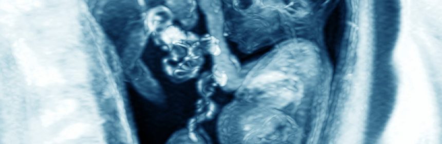
Some scientists are betting on magnetic resonance imaging scans, or M.R.I.s, as the most sensitive detectors of placental problems. They are using a method that measures oxygen levels in the blood; it is quick and, so far, seems to catch problems as early as the second trimester. Several teams worldwide are evaluating the technique, each in hundreds of women.
But M.R.I. is not widely in use in obstetricians’ offices, Dr. Abuhamad said, in contrast with ultrasound machines, which would be a more practical option. Traditional ultrasounds can show the structure and location of the placenta, not how well the organ is functioning. But advances over the past five years have sharpened the machine’s focus. One enables the device to detect tiny blood vessels; another, called elastography, was developed to examine the liver, and can help measure the density of placental tissue.
Dr. Abuhamad’s team is using these advances in ultrasound to chart placental health in about 500 pregnant women, including 300 at high risk of complications. They are collecting ultrasound data and blood samples from the women at eight time points during pregnancy to see which early features track with problems later on.
Other teams are trying to identify particles the placenta may release into the bloodstream because that could lead to a simple blood test for diagnosing problems. And one group of researchers is developing an oximeter, a device that quantifies the light reflected back through layers of fat as a measure of blood oxygen.
It will be at least five years before any of these tests makes their way to doctors’ offices. But when they are ready, they are likely to have a huge impact on obstetric practice, said Dr. Diana W. Bianchi, director of the National Institute of Child Health and Human Development.
“The way that prenatal care is currently structured, you hardly see your obstetrician in the first trimester,” she said. “And by the time you get to the third trimester, you’re seeing the obstetrician weekly.”
Instead, when placental screening identified a problem, women might be encouraged to see their doctors frequently through the first trimester. “Knowing that this starts early in the first trimester,” said Dr. Abuhamad, “could we then intervene in the first trimester — identify early, intervene early and prevent the complications?”
Source: Read Full Article