Reinventing the Uterus, One Organoid at a Time
CAMBRIDGE, Mass. — Most labs devoted to women’s diseases are accompanied by obvious symbols of womanhood: a rose, a tulip, an hourglass silhouette. Not Linda Griffith’s. Tucked away in the building for biological engineering, the M.I.T. Center for Gynepathology Research is marked only by the letters CGR in red and black, the G formed from a curved arrow representing the hand of the engineer.
“We needed something that wasn’t all pink and flowers,” said Dr. Griffith, the lab’s director. “We really thought it should be, like, ‘This is science.’”
Dr. Griffith founded the lab in 2009 with the goal of helping researchers solve endometriosis, a chronic disorder in which tissue similar to that which normally lines the uterus instead grows outside it. The disease strikes one in 10 women, as well as trans men and nonbinary people who menstruate. Its hallmarks are extreme pain and, in some cases, infertility.
Yet it suffers from a branding problem: It falls into the abyss of “women’s diseases” (overlooked), diseases that don’t kill you (unimportant) and menstrual problems (taboo). Researchers often call endometriosis “benign,” as in noncancerous — but doing so, Dr. Griffith believes, lessens the seriousness of a common, painful disease.
Her mission is to change the conversation, from one of women’s pain to one of biomarkers, genetics and molecular networks. “I don’t want to make endometriosis a women’s issue,” she told the M.I.T. Tech Review in 2014. “I want to make it an M.I.T. issue.”
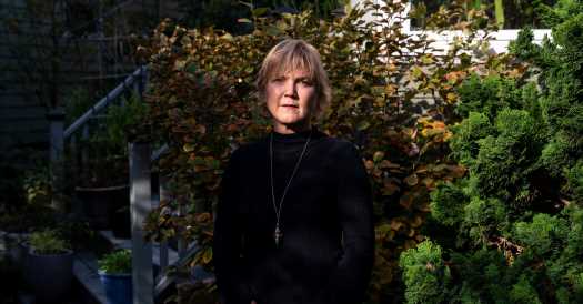
Dr. Griffith, spry at 60, with a fringe of blonde hair and a slight Georgia drawl, is uniquely poised to help effect that transition. She started her career in bioengineering, sculpting organs like liver and bone from scratch by seeding polymer scaffolds with living cells. In 1997, she helped create an iconic creature called the “earmouse” by injecting a human ear-shaped scaffold with cartilage from a cow’s knee, and growing it on the back of a lab mouse.
Now she brings those skills to the task of better understanding the uterus. In her lab, she has begun growing uterine organoids — tiny domed droplets, with glands that look like swirling craters — from the uterine cells of endometriosis patients. These “patient avatars” are ideal tools for testing potential new treatments for the disease: Biologically, they are closer to human uterine cells than those of mice (which don’t naturally menstruate). And they enable researchers to sidestep the ethical issues that would arise with human trials.
“That’s really the power of this,” Dr. Griffith said. “You can take patients who we know how they respond or do not respond to therapies, and compare and start to understand and tease apart why that is.”
Her research highlights what a remarkable organ the uterus truly is — and not just during its signature function, pregnancy. Humans, unlike almost every other mammal, shed their entire endometrium — the womb’s inner lining — once a month and grow it back, whether or not a fertilized egg takes hold.
Dynamic, resilient and prone to reinvention, the uterus offers a window into some of biology’s greatest secrets: tissue regeneration, scarless wound healing and immune function. “The endometrium is inherently regenerative,” Dr. Griffith said. “So studying it, you’re studying a regenerative process — and how it goes wrong, in cases.”
Now, her work “is drawing the interest of those who have never worked on or never thought about endometriosis,” said Dr. Stacey Missmer, a reproductive biologist at Michigan State University and co-director of the Boston Center for Endometriosis. Essentially, Dr. Missmer said, Dr. Griffith is saying: “All you cool kids in the other disciplines, this is a really interesting area to ask questions.”
A name for the pain

Before Dr. Griffith turned her scientific lens on the womb, she spent years trying to avoid thinking about the pain it caused her. For nearly three decades doctors dismissed her symptoms — stomach-turning nausea, stabbing pelvic pain and alarming levels of blood loss during her period — as just part of being a woman.
“I felt like I was being gaslighted,” she said.
She grew up fearless, a tree-climbing Girl Scout in Valdosta, Ga. In high school, she sewed her own clothes, earned a black belt in karate and fixed her family’s car radiator. “There was nothing we couldn’t do, whether we were male, female, whatever,” said her younger sister, Susan Berthelot. “We had a lot of confidence, and a lot of love, and a lot of freedom.”
But when Dr. Griffith hit puberty, her body began imposing limitations. Her period was so agonizing it would leave her curled in the fetal position for days. When she was 13, a gynecologist prescribed birth control pills, a scandalous proposition. “In the South especially, it was not done,” she said. Her mother, at a loss, gave her gin.
Unable to control what was going on inside her body, she focused on what she could control: math, and building things. She went to Georgia Tech on a scholarship to study chemical engineering. But she found herself failing tests when she was on her period, and going to the infirmary to get monthly shots of the opioid Demerol.
By the time she began graduate studies at the University of California, Berkeley, she had developed an elaborate period regimen: She wore all-black outfits, inserted three Super Plus tampons and swallowed upward of 30 Advil tablets a day. But her pain kept increasing. When she consulted a male doctor, he took one look at her black leather jacket, pixie cut and Bugatti motorcycle and diagnosed her as “rejecting her femininity.”
Her real diagnosis came by accident. In November 1988, soon after she went to M.I.T. as a postdoc, she checked into the Brigham and Women’s Hospital in Boston to drain a small cyst on her left ovary. She woke up the next day to find a row of staples along her midriff, holding together a six-inch incision.
Her gynecologist told her she had a disease called endometriosis, which had fused her pelvic organs together with a sticky, speckled tissue that resembled the lining of her uterus. This rogue tissue responded to her monthly hormone cycle, swelling, shedding and attempting to bleed; that was the origin of the pain.
Surgeons had burned or scraped off as much of the tissue as possible; there was little else they could do. In 1940, the gynecologist who gave endometriosis its name, Dr. John Sampson, deemed the disease “tantalizingly alluring and elusive.” A half-century later, not much had changed. There was no cure, and researchers still didn’t know exactly how endometriosis took root.
Still, Dr. Griffith treated the diagnosis as good news. “To have someone tell me something was wrong with me, it was a huge relief,” she said.
Her gynecologist presented two options: She could go on Danazol, a hormone-blocking drug that would halt the growth of the disease but would also send her body into a menopause-like state; or she could get pregnant, a common recommendation in the 1980s, and not uncommon today.
The medical reasoning — which has since been questioned — was that by temporarily stopping menstruation, pregnancy could reduce symptoms and slow or reverse the growth of lesions. “It was almost viewed as a two-for-one benefit,” said Dr. Elizabeth Stewart, who performed Dr. Griffith’s first surgery. “It’s clear there was some sexism in the approach to endometriosis then. I think there’s still some now.”
Dr. Griffith recalls her then-husband answering for her: “We’ll have a baby.”
She opted for the Danazol. Eight years later, she divorced the husband.
Soon she was jump-starting the field of biological engineering, developing technologies to 3D-print organ scaffolds and growing artificial human ears on the backs of mice. She was an architect; her medium was the building blocks of life. But it never occurred to her to try to solve her own disease.
“Psychologically, it wasn’t something I wanted to think about,” she said. “I just wanted to pretend like it wasn’t happening.”
A ‘women’s thing’ worth doing
The turning point came in 2007, when a member of M.I.T.’s board of trustees, Susan Whitehead, asked her to speak at a Women in Science and Engineering luncheon about how her work on tissue engineering could benefit women.
Dr. Griffith was annoyed. “I was working on all the things that guys were working on,” she later recalled at a 2018 scientific meeting. “It didn’t ever occur to me to work on a women’s thing.” But Whitehead was a friend, so she agreed.
Near the end of the event, the moderator asked her where she saw herself in 10 years. Something welled up inside her. She had just had her eighth surgery for endometriosis, and had helped her 16-year-old niece, Caitlin, receive a diagnosis for endometriosis after years of having doctors attribute her symptoms to stress. Watching Caitlin go through the same ordeal “made lava shoot out of my head,” Dr. Griffith recalled recently.
“I have a chronic disease called endometriosis,” she blurted out to the luncheon audience, and mentioned her niece. “There’s no better treatment for her, 30 years younger than me, than there was for me when I was 16.” If a major breakthrough in treatment didn’t come soon, “that’s where I’m going to be in 10 years,” she said. “Maybe it’ll be solved, but I don’t think so.”
The audience broke into applause.
Dr. Griffith reset her goals. When it came to making liver and bone, “so many other people could do them,” she recalled. “But there was this one thing only I could do.” She had recently been awarded a MacArthur “genius” grant, which came with $500,000 for any research project.
In 2009, she used it toward opening the Center for Gynepathology Research, the only engineering lab in the nation to focus on endometriosis. (In October 2020, federal funding for endometriosis research doubled from $13 million to $26 million after Representative Abby Finkenauer of Iowa, 32, shared her own endometriosis journey on the House floor.)
At the launch event for the center, Padma Lakshmi, host of Top Chef and co-founder of the Endometriosis Foundation of America, lamented the lack of research on such a devastating disease.
“I have to say, I’m really shocked that it’s the first research center of its kind in America,” she said. “That is stunningly bad news on the one hand, that she’s the first one doing it. On the other hand, better late than never. Thank God for Dr. Linda Griffith.”
A window into the womb
Imagine the uterus as an orange, with the lining as the rind: fluffy, living tissue that serves as a plush bedding for a potential embryo. Each month, triggered by a drop in the hormone progesterone, the lining sloughs off and grows anew, complete with delicate, spiraling blood vessels.
This process repeats itself swiftly, scarlessly, without a trace of injury, again and again, as many as 500 times in a woman’s life. “How the body can coordinate that is extraordinary,” said Dr. Hilary Critchley, a reproductive biologist at the University of Edinburgh — and still poorly understood.
But this remarkable dynamism, some researchers argue, is a double-edged sword. “A little thing gets out of balance, and there you go,” said Dr. Griffith.
Dr. Griffith’s models offer a glimpse into what happens when the process goes wrong — for instance, when this growing tissue takes root in places it shouldn’t. Her bits of bioengineered tissue allow researchers to visualize the growth of lesions and systematically parse the role of immune cells, inflammation and hormones in the disease.
“You’re actually seeing in three dimensions what’s going on inside the uterus and this gland formation and nerve formation,” said endometriosis specialist Dr. Keith Isaacson, who co-directs her lab. “That is incredibly exciting.” (Dr. Isaacson, who has been Dr. Griffith’s endometriosis surgeon since 2000, and provides the patients’ cells for her models.)
With her background in systems engineering, Dr. Griffith sees the uterus not as an island but as an organ that interacts intimately with everything around it. To capture these systemic interactions, her team connects her models to other organs like bone marrow, gut and liver, and hopes one day to seed them with blood vessels, nerve cells and immune cells.
The insights from this research transcend the womb. For instance, one enduring mystery about the disease is how lesions can appear in places as far-flung as the lungs, eyes, spine and even the brain. Dr. Hugh Taylor, chair of the department of obstetrics, gynecology and reproductive sciences at Yale School of Medicine, is investigating whether stem cells, which are plentiful in the uterine lining, could contribute to this process by circulating throughout the body.
Because uterine stem cells are relatively accessible, they could also be a boon to regenerative medicine. Dr. Taylor has shown that, like other stem cells, they can be grown in vitro into new neurons and insulin-making cells to treat diseases like Parkinson’s and diabetes.
Another area ripe for improvement is diagnosis. One of the most frustrating aspects of endometriosis is that women typically wait seven to 10 years or more to learn that they have the disease, a process that requires invasive surgery. Now, researchers are developing a simple test to screen for genetic markers of endometriosis in menstrual blood and return a near-instant diagnosis.
Just a few milliliters of this blood, collected on a sponge, provides a wealth of markers of health and disease, said Christine Metz, an immunologist at Northwestern’s Feinstein Institute who is developing the test with Peter Gregersen, a geneticist there. Besides endometriosis, it could also help doctors screen for pelvic inflammation, infertility, fibroids, environmental toxins and early cancer.
“We were kind of surprised that it had been neglected as a natural resource,” Dr. Metz said.
They soon discovered why. In 2018, Dr. Metz began approaching male gynecologists to ask their patients for menstrual samples, and “some people said we were completely insane,” she recalled. Others, she added, “had never heard of a menstrual cup. Which was also, I’m going to say, disappointing.” They began asking patients directly for samples, and have since enrolled 1,000 patients in their study.
One might well ask why more researchers have not focused on the uterus until recently. Bioengineers in particular have always taken an interest in tissues that regenerate and self-heal. “And yet it took them how many decades to recognize that one of the most regenerative tissues is found inside the uterus?” asked Kathryn Clancy, a biological anthropologist who studies reproduction at the University of Illinois.
The reason, she believes, is simple: “Because none of the researchers had uteruses.”
Lessons from cancer
Three stone cherubs form an arc above the doorway to Dr. Griffith’s kitchen in Cambridge, Mass. They were a gift from her mother, to commemorate the embryos that she and her current husband, Doug Lauffenburger, a systems biologist with whom she shares a lab at M.I.T., made in 1997 through in vitro fertilization. Endometriosis prevented the embryos from implanting.
She held on to her dream of having children, but in 2001, just after her 40th birthday, the pain in her abdomen grew unbearable. On Sept. 11, as the Twin Towers fell, she rushed to the hospital in a fog of painkillers and underwent a hysterectomy with Dr. Isaacson. (Endometriosis pain is the leading cause of hysterectomies for American women in their 30s.)
“There was no decision,” Dr. Griffith recalled. “It was hysterectomy or death.”
Even after that, her disease returned, twice. Then in 2009, just after she had pivoted to studying endometriosis, she faced a new obstacle: cancer.
Dr. Griffith likes to say that compared to endometriosis, stage 4 breast cancer was a walk in the park. “Not like a super-beautiful day — like a stormy-day walk in the park,” she added. “But it was, like, people understood.” Colleagues wrote her cards, sent her food, extended condolences. Her dean offered her a sabbatical semester.
Dr. Griffith soon learned that the way breast cancer research was categorized was far ahead of endometriosis. Doctors used molecular tests to classify patients into subtypes, which dictated which targeted treatment they should receive. With endometriosis, “there’s no metrics,” she said. “This was this huge thing for me that was so crystallizing.”
Dr. Griffith knew that her disease, like cancer, was not one disease but many, a medusa of waving tentacles. She began talking to Dr. Lauffenburger, who had been studying breast cancer for over a decade, about how to take a similar approach to classifying endometriosis patients.
Together, they identified networks of inflammatory markers that tended to be associated with more painful manifestations of the disease and fertility, and published their findings in Science Translational Medicines in 2014. The work was cited as the first step toward creating subtypes of the disease. “That was really us together, because it was his vision of systems biology but filtered through my practical connection to the clinic,” Dr. Griffith said.
For the next year, she held lab meetings from her hospital bed in between chemotherapy sessions. “We transformed our lab meetings, literally,” said Dr. Nicole Doyle, a postdoctoral fellow in Dr. Griffith’s lab at the time. “We just showed up for her chemo treatments and would sit there with her. That diagnosis had to adapt to her life, not the other way around.”
Throughout chemo, Dr. Griffith never seemed to waver in her positivity. When she shaved off her hair, she threw a lab party. But Dr. Lauffenburger found it excruciating to watch his wife suffer from this new foe, after battling the old one for so long.
When it came to cancer, “I viewed it as a terrible thing,” he said one evening over dinner at their home.
Dr. Griffith saw it differently: She took a curse and turned it into a gift.
“It was a terrible thing,” she allowed. “But it was a good thing, scientifically.”
[Like the Science Times page on Facebook. | Sign up for the Science Times newsletter.]
Source: Read Full Article