Treatment Options for Outer Ear Cancer

Generally, ear cancer is defined as the uncontrolled development and growth of cancer cells in the ear. Ear cancer is comparatively the lesser prevalent of all types of cancers. Typically, cancer in the ear develops as a skin cancer in or around the external portion of the ear. Cancer cells might grow in any of the three parts of the external ear constituting the fleshy visible pinna (the auricle), the ear canal, and the outer layer of the eardrum (tympanic membrane).
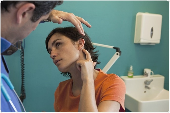
Credit: Image Point Fr
Primary Treatment Options for Outer Ear Cancer
Choices of remedial measures include surgical operation or radiation therapy, or both. These remedial measures almost cure cancers in the external portion of the ear. The category and extent of surgery to be performed are based on the stage of the cancer. The most common types of surgery are listed below:
- Mohs micrographic surgery
- Wide Local Excision
- Excisional biopsy
- Lymph node dissection
- Otoplasty
Mohs Micrographic Surgery
Mohs micrographic surgery is a procedure in which thin layers of cancer-containing skin are progressively removed along with the surrounding thin layer of healthy tissues. The thin layer of cancer-containing skin, which has been removed through Mohs surgery, is subjected to sectioning, freezing, staining, and mapping in detail, and then it is completely examined under a microscope.
If cancer is still found in the inner or outer layer of this removed surrounding thin layer of tissue, the surgeon removes the indicated cancerous tissue from the patient through Mohs procedure. This procedure is repeated on any locations still containing cancer cells until no further cancer cell is found under a microscope.
Wide Local Excision
In addition to the initial biopsy, wide local excision is used in melanomas (malignant tumors in skin cancer) that tend to recur.
The procedure generally excises 1 to 2 cm of healthy skin layer around the melanoma location; nevertheless, variation in this margin depends on factors such as the impact of tumor depth and its extent of spread into the dermis and hypodermis layers of the skin. Closing the wide local excision with stitches is often performed on smaller lesions, but skin grafts or skin flaps may be employed on larger lesions.
Excision Biopsy
In an excision biopsy approach, the surgeon uses a lancet to completely excise the entire tumor-affected region and excision margin of healthy skin around it. The surgical wound around the tumor site may be stitched up.
The specimen of removed tissue is observed under a microscope to make sure that all tumor cells have been excised. This procedure may be repeated later until no further cancer cell is examined on a skin specimen under a microscope. The highest overall cure rate of primary cancers by this approach is about 92%.
Lymph Node Surgery
In lymph node biopsy, lymph node tissue is excised and examined under a microscope to identify the symptoms of a tumor or other infection. Lymph nodes are listed under the human immune system. Lymph nodes are present everywhere across the body, but swollen lymph nodes in the neck, armpits, and groin area only can be easily identified.
Enlargement of lymph nodes usually denotes the symptom of cancer or other infections. No specific care is required for most of the lymph nodes in cases where they get enlarged owing to local infection or trauma.
Otoplasty or Ear Reconstruction Surgery
Ear reconstruction remains one of the most difficult areas of plastic surgery. The first step in reconstructive surgery is to develop a skin pocket at the ear location by re-orientating the tissue available and skin thinning to match the cosmetics of the adjacent ear skin.
Three small pieces of rib cartilage are harvested. The rib cartilage is sculpted and cabled together with fine wire made of stainless steel to make a detailed framework that resembles an ear. The whole framework is then introduced inside a pocket. Gentle suction may be feasible in fusing the skin and the ear structure together.
The second step involves positioning of the new ear in such a way that it normally projects out from the head like a normal ear. The ear is lifted from its bed and positioning is maintained by another piece of cartilage that is fixed behind the ear. To end the process, a layer of tissue and skin graft are taken and placed over the uncovered cartilage.
Radiotherapy
In radiotherapy, very intensive energy (radiation) rays are used to treat cancer. To start with radiation treatment, the patient has to consult with the radiologist who designs the treatment schedule, which can last between 4 and 6 weeks.
If the cancer on the exterior portion of the ear or pinna is small, then radiation therapy is enough to kill the cancer cells. It is also possible to have radiotherapy post-operation in situations when the physician is unable to dissect a clear margin around the cancer. This reduces the risk of cancer recurrence after removal of tumor through surgery.
Sources:
- http://www.cancernews.com.au/cancer-of-the-ear/
- www.cancerresearchuk.org/…/outer-treatment
- www.skincancer.org/…/scc-treatment-options
- https://www.melanoma.org.au/understanding-melanoma/treatment-options/
- www.skincancer.org/…/scc-treatment-options
- medicine.yale.edu/…/lymph.aspx
- www.royalfree.nhs.uk/…/
- www.cancerresearchuk.org/…/outer-treatment
Further Reading
- All Ear Content
- How Does the Ear Work?
- Causes of Hearing Loss
- Causes of Tinnitus
- Earwax – Cerumen
Last Updated: Feb 26, 2019
Source: Read Full Article