What is Mitochondrial Fractionation?

Mitochondrial fractionation can be used to obtain isolated mitochondrial fractions from living cells.
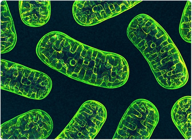
Image Credit: 3d_man/Shutterstock.com
These fractions are samples of concentrated mitochondrial material that can be used to study the biochemical processes of the mitochondria in the sample. Mitochondrial Fractionation can be used for either research or diagnostic use.
The assay is used to investigate apoptotic and signal transduction pathways. The separated fractions it produces can also be further investigated by Western blotting, ELISA, or other assays, to uncover translocation of factors between two fractions.
The basis of the method itself involves breaking down a sample of cell cultures, mechanically or chemically, and then separating the mitochondrial fraction from other cell material via centrifugation.
It has become a firmly established method in biological laboratories due to its robustness, simplicity, and ease of use, and that it requires no ultra-centrifugations or toxic chemicals.
Assay protocol
There are several stages to the standard protocol for conducting mitochondrial fractionation. The first step involves collecting the cell sample by centrifugation, washing them, and resuspending them with a buffer containing DTT and Protease Inhibitors.
After a brief incubation period on ice, the cells can then be homogenized using a tissue grinder, the efficiency of which should be checked by observation of the homogenized suspension under a microscope.
Excessive homogenization should be avoided to prevent damage to the mitochondrial membrane. Following this step, the homogenate can be transferred into a microcentrifuge tube and centrifuged again.
Then, the supernatant is collected and transferred into a fresh tube and centrifuged again. The pellets from the second centrifuge should be saved and resuspended if intact mitochondria are required.
Various labs manufacture mitochondrial fractionation assays that will vary slightly in their protocol, however, the method always involves using centrifuges to separate mitochondria from cell cultures that have been broken down chemically or mechanically.
Why study mitochondrial fractions
The study of mitochondria is vital to numerous areas of biology. Because the organelles are responsible for a variety of essential functions, such as amino acid metabolism, carbohydrate metabolism, fatty acid oxidation, programmed cell death (apoptosis), free radical formation, signal transduction, as well as host genes encoding several important proteins.
Investigating and studying mitochondria is therefore fundamental to developing knowledge around cell function, as well as to grow an understanding of mitochondrial disorders and mitochondrial responses to pharmacological agents.
Defective mitochondrial function can lead to mitochondrial disorders and is also linked with various diseases, making mitochondrial fractions valuable to the development and testing of new therapeutic interventions.
Mitochondrial fractions are the gateway to understanding the activity of mitochondria because methods that use whole organism or whole tissue extractions can be hindered in producing reliable and accurate data of mitochondrial metabolism because of the disruption caused by the presence of other cytosolic metabolites.
Mitochondria’s role in disease
The activity of mitochondria has been linked with several diseases, therefore, the study of them is significant in growing an understanding of the development and progression of the disease, and even to the development of new treatments.
Mitochondrial DNA (mtDNA) mutations have already been linked with neuromuscular and neurodegenerative mitochondriopathies, diabetes, cardiovascular diseases, gastrointestinal disorders, skin disorders, aging, and cancer.
For example, a growing number of studies are exploring the role of mitochondria in cancer. Mutations in mtDNA have been reported in renal adenocarcinoma, colon cancer cells, head and neck tumors, astrocytic tumors, thyroid tumors, breast tumors, ovarian tumors, prostate and bladder cancer, neuroblastomas and oncocytomas.
Mutations in nuclear DNA (nDNA) have been found to indirectly affect mtDNA, and have also been linked with specific cancers.
Also, mitochondrial activity is known to be essential to maintaining the proper function of tissues and organs that rely on high and consistent levels of energy, such as the heart. Disturbances to mitochondria have therefore been linked with cardiovascular disease (CVD), and the use of mitochondrial fraction is being used in studies to investigate this link and elucidate the full role of mitochondria in CVD.
As well as being implicated in various diseases, there is a specific group of inherited disorders that are classified through the failure of mitochondria to produce enough energy to allow the body to function properly.
These are referred to as mitochondrial diseases. They can affect any part of the body, from brain cells and nerves to the muscles, heart, liver, kidneys, pancreas and even eyes and ears. While these diseases are rare, around 1,000 to 4,000 children are born in the US each year with the disease, which can be fatal, and for which there is no known cure.
Therefore, the use of mitochondrial fractionation to study the behavior of mitochondria in these illnesses is essential to the continued investigation of new potential therapies that could improve the quality of life and prevent fatalities.
Summary
Mitochondrial fractionation is a vital method for separating cultures of mitochondria from cell samples in order to study them. This technique has proved invaluable to growing an understanding of the function of mitochondria and their role in a number of diseases, as well as helping to develop potential new therapies for disease.
Sources
- Chistiakov, D., Shkurat, T., Melnichenko, A., Grechko, A. and Orekhov, A. (2017). The role of mitochondrial dysfunction in cardiovascular disease: a brief review. Annals of Medicine, 50(2), pp.121-127. https://www.ncbi.nlm.nih.gov/pubmed/29237304
- Villa-Cuesta, E. and Rand, D. (2015). Preparation of Mitochondrial Enriched Fractions for Metabolic Analysis in <em>Drosophila</em>. Journal of Visualized Experiments, (103). https://www.ncbi.nlm.nih.gov/pmc/articles/PMC4845975/
- Wallace, D. (2012). Mitochondria and cancer. Nature Reviews Cancer, 12(10), pp.685-698. https://www.ncbi.nlm.nih.gov/pmc/articles/PMC4371788/
Further Reading
- All Mitochondria Content
- What is the Electron Transport Chain?
- Analyzing Mitohormesis
- Mitochondrial DNA Organization and Replication Steps
- Mammalian Mitochondrial DNA (mtDNA) Transcription
Last Updated: Feb 7, 2020

Written by
Sarah Moore
After studying Psychology and then Neuroscience, Sarah quickly found her enjoyment for researching and writing research papers; turning to a passion to connect ideas with people through writing.
Source: Read Full Article