New noninvasive optical imaging approach for monitoring brain health in traumatic brain injury patients

Prof. Mohammed N. Islam leads a team of researchers who have developed a new cost-effective, portable, non-invasive means of monitoring cerebral, tissue, and organ metabolism and hemodynamics simultaneously. The tool, a noninvasive Super-Continuum Infrared Spectroscopy of Cytochrome C-Oxidase (SCISCCO) system, can aid the early detection of brain injury or neuronal dysfunction and can continually monitor brain health to help guide therapies and treatments for injury.
“Internal bleeding due to traumatic injuries is a major cause of preventable deaths,” Islam said. “Our noninvasive method for monitoring tissue metabolism could help improve the diagnosis and monitoring of conditions such as concussions, stroke, traumatic brain injury, and other medical conditions.”
Traumatic brain injury (TBI) results in structural damage to the brain. Patients require immediate medical intervention, evacuation, and intensive care, and many will need life-long assistance after recovery. While TBI can happen to anyone, it is known as the signature wound for veterans of the wars in Iraq and Afghanistan. The Department of Defense reports that over 320,000 troops suffered TBIs from 2010 to 2015. However, the military still has no objective way of diagnosing it in the field.
In addition, three-quarters of traumatic brain injury survivors experience a mild traumatic brain injury (mTBI), or concussion. Concussion is challenging to diagnose, for it involves a brief change in mental status without any observable structural damage to the brain. However, individuals with a concussion may still suffer from life-long cognitive or psychological challenges, such as memory problems and post-traumatic stress disorder.
Current methods to monitor changes in blood flow to the brain rely on hemodynamic neuroimaging systems, but these systems can’t detect changes in the neural tissue itself. This can result in illness or injury going undetected and, therefore, untreated. To address this issue, Islam’s team focused on measuring the optically active markers of cellular function, such as cytochromes.
Cytochrome C-Oxidase (CCO) is a photo-sensitive enzyme that is responsible for more than 95% of oxygen metabolism in the body. CCO concentration is also significantly higher in the brain than in extra-cerebral tissues. Therefore, neural metabolism could be measured by capturing changes in CCO, and simultaneous measurements of CCO and hemoglobin redox states could provide additional information on the brain’s metabolism and hemodynamics, which could aid in the diagnosis and management of neurological injury or illness.
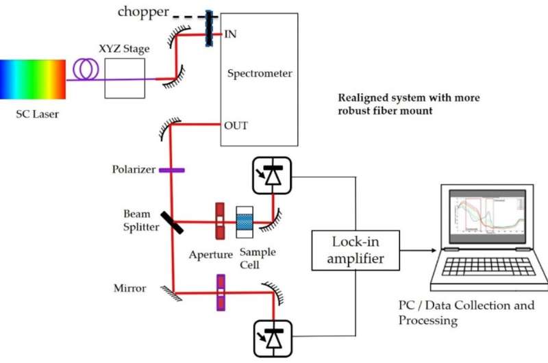
The conventional means of measuring CCO relies on lamp-based light sources to provide the illumination, but these systems are plagued with generally poor signal-to-noise ratio. Islam’s team, by contrast, uses an all fiber integrated super-continuum light source for a noninvasive optical imaging approach. This approach greatly improves signal-to-noiseratios, and results in more definitive measurements of CCO. The result is the Super-Continuum Infrared Spectroscopy of Cytochrome C-Oxidase (SCISCCO) system.
“Our system can provide almost an order of magnitude improvement in brightness compared with the tungsten-halogen lamps typically employed for CCO measurements,” Islam said.
The SCISCCO system is also extremely versatile.
“The SCISCCO system could be applied to range of uses, from serving as a new tool for screening concussion patients—such as being used in the cognitive attention test results—to use in an intensive care unit to gauge a patient’s organ response to treatments,” Islam said.
Specifically, it can perform measurements of CCO, oxygenated hemoglobin (HbO), and deoxygenated hemoglobin (HbR) to determine the metabolic function of any organ, such as the liver, kidney, heart, and lungs. It can also be used to measure and monitor the metabolic function of tissues, such as muscle tissue, nervous tissue, and epithelial tissues.
Islam’s team is currently performing human studies in an intensive care unit (ICU) setting with the U-M Neurosurgery department.
Source: Read Full Article
