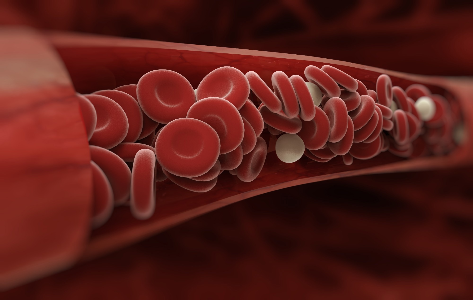
Investigating the relationship between red blood cells and the clinical course of COVID-19 in children

In a recent study published in Scientific Reports, researchers performed the ImmunCoviDD19-study among school students from urban Dresden and rural Saxony in Germany between June 24 and July 16, 2021.
The study investigated morphological and mechanical characteristics of red blood cells (RBCs) after severe acute respiratory syndrome coronavirus 2 (SARS-CoV-2) infection.

Introduction
They enrolled 131 students in this study. However, they could successfully perform real-time deformability-cytometry (RT-DC) analysis for only 121 children and adolescents from 14 secondary schools to understand the relationship between RBC deformation and the pathophysiology of coronavirus disease 2019 (COVID-19).
Background
RBCs are one of the key players in microcirculation, hypoxemia, and tissue oxygenation under physiological conditions. Severe COVID-19 triggers microcirculation as a compensatory response.
Consequently, several studies have described changes in the number and morphology of RBCs during COVID-19. For instance, Nader et al. described an increased RBC aggregation in severe COVID-19.
Likewise, Piagnerelli et al. found that RBCs deformability remained unaltered in patients with acute respiratory distress syndrome (ARDS), which led to severe hypoxemia in contrast to patients with bacterial sepsis, thus, explaining the intact microcirculation observed during severe COVID-19.
Other studies have shown a correlation between RBC parameters, mean corpuscular volume (MCV), mean corpuscular hemoglobin (MCH), and RBC distribution width (RDW) and COVID-19 severity, which could be a potential prognostic marker depicting the clinical course of COVID-19.
Overall, COVID-19-induced changes in RBC properties might impact microcirculation, heightening the risk of pulmonary embolism and micro thrombosis.
About the study
In the present study, researchers used retrospective data from the SchoolCoviDD19, a survey-based study that allowed exploratory subgroup analyses of all COVID-19 seropositive participants concerning seroconversion time.
The SchoolCoviDD19-study categorized participants who tested polymerase chain reaction (PCR) positive on or before December 10, 2020, as seropositive with a time of seroconversion >six months. The others were seronegative with a time of seroconversion <six months. Moreover, this study only included subjects who were fully COVID-19 vaccinated.
Further, the researchers analyzed the median and interquartile range (IQR) of area, deformation, and standard deviation (SD) of brightness for RBCs. Furthermore, they compared the data between SARS-CoV-2-seropositive, seronegative, and vaccinated participants by two-tailed Welch’s t-tests.
Finally, they performed partial correlations between either group by SARS-CoV-2-serostatus by adding age and gender as confounding variables. They controlled type 1 errors and adjusted p-values using Holm-Bonferroni correction for multiple comparisons.
Results
The RT-DC analysis in this study encompassed 121 participants with an average age of 14.93 years, of which 63 and 49 subjects were seropositive and seronegative for SARS-CoV-2 immunoglobulins G, respectively, with no age- and gender-based variations across both groups.
In addition, nine participants reported a complete COVID-19 vaccination status at the time of blood collection and were older than SARS-CoV-2 seronegative participants.
The authors noted significant RBC alterations post-COVID-19 in children and adolescents stratified by time since SARS-CoV-2 seroconversion. In SARS-CoV-2-seropositive participants, average RBC deformation was markedly higher, even after controlling for sex and age.
The IQR of RBC deformation tended to be lower but did not withstand the Holm-Bonferroni correction. However, the median RBC area was similar in SARS-CoV-2-seropositive and seronegative participants.
RBC deformability depends on the cytoskeletal integrity. Thus, increased RBC deformation likely indicated fluidization of the cell membrane in COVID-19 patients. Studies identified membrane-bound protein band-3 as the target of SARS-CoV-2 during the invasion of RBCs.
Its fragmentation, thus, also explains a loss of membrane integrity and enhanced RBC deformation.
An immune complex deposition on the RBC membrane is another RBC modulation observed in COVID-19 patients. In many cases, mechanisms causing an increased RBC deformation might be physiological compensations to prevent COVID-19-related micro thrombosis and hypoxemia.
Intriguingly, fully vaccinated participants had increased RBC deformation relative to seronegative participants, further favoring the notion that RBC deformation might be a direct consequence of the human immune response against SARS-CoV-2 spike presentation, though this observation warrants further validation.
Another remarkable study finding was the increased SD of RBCs brightness post-SARS-CoV-2 infection, a parameter that reflects structural alterations in the cells due to direct invasion of RBCs.
RBCs have a lifespan of ~120 days, so most likely, resuming RBC deformation to levels of seronegative participants after six months is a physiological recovery process post-acute SARS-CoV-2 infection, as assessed in a sub-analysis based on the time of seroconversion.
Conclusions
Overall, the study findings suggested that SARS-CoV-2 infection in children (also adolescents) increased RBC deformation during the initial six months, representing a compensatory mechanism that prevented severe pathological changes and symptoms during acute COVID-19.
In the future, RBC deformation in children and adolescents, assessed via RT-DC, could potentially be used as an easy-to-use point-of-care test in clinical diagnostics.
-
Eder, J. et al. (2023) "Increased red blood cell deformation in children and adolescents after SARS-CoV-2 infection", Scientific Reports, 13(1). doi: 10.1038/s41598-023-35692-6. https://www.nature.com/articles/s41598-023-35692-6
Posted in: Child Health News | Medical Science News | Medical Research News | Medical Condition News | Disease/Infection News
Tags: Acute Respiratory Distress Syndrome, Adolescents, Blood, Cell, Cell Membrane, Children, Clinical Diagnostics, Coronavirus, Coronavirus Disease COVID-19, covid-19, Cytometry, Diagnostics, Embolism, Hemoglobin, Hypoxemia, Immune Response, Membrane, micro, Morphology, Pathophysiology, Polymerase, Polymerase Chain Reaction, Protein, Pulmonary Embolism, Red Blood Cells, Respiratory, SARS, SARS-CoV-2, Sepsis, Severe Acute Respiratory, Severe Acute Respiratory Syndrome, students, Syndrome, Thrombosis

Written by
Neha Mathur
Neha is a digital marketing professional based in Gurugram, India. She has a Master’s degree from the University of Rajasthan with a specialization in Biotechnology in 2008. She has experience in pre-clinical research as part of her research project in The Department of Toxicology at the prestigious Central Drug Research Institute (CDRI), Lucknow, India. She also holds a certification in C++ programming.
Source: Read Full Article