G Protein-Coupled Receptors (GPCRs) Overview

By Jeyashree Sundaram, MBA
G protein-coupled receptors (GPCR), G-protein linked receptors, serpentine receptors, or heptahelical receptors are the largest superfamily of membrane proteins in the human genome.
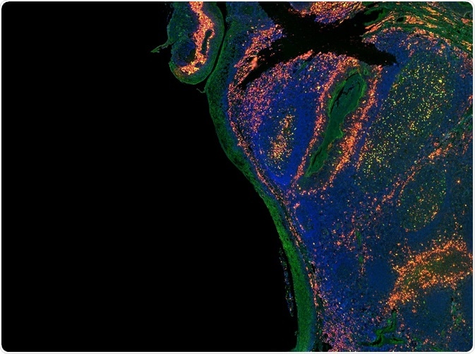
Credit: Carl Dupont/Shutterstock.com
GPCRs possess a heptahelical structure and support normal transmembrane cell signaling processes for growth and cellular development. They are mainly found in eukaryotes but have also been observed in some prokaryotes.
GPCRs are primarily involved in initiating the sensory and regulatory mechanism of cells, acting as receptors for internal actuators and external ligands, respectively.
Various physiological functions, including the development of immune systems and cell metabolism, necessitate the use of GPCRs. For this reason, they also act as drug targets inthe treatment of psychological and mental disorders.
Structure
GPCRs are the largest superfamily of protein receptors found in the human genome. They are protein receptors that possess a seven alpha-helical transmembrane core with a carboxyl-terminus (C-terminus) and an amino-terminus (N-terminus) in the interior and exterior of the membrane, respectively.
The human genome encodes around 800 individual G coupled protein receptors, of which the olfactory receptors constitute a major segment (nearly 460 GPCRs).
Classification
The protein receptors are briefly classified into six families, based on the similarity in their sequential arrangement:
Class A (the rhodopsin family):
The rhodopsin family consists of 701 members (about 80% of the overall species). The structural elements of this family are mostly incorporated within the alpha group. The beta, gamma, and delta forms are found only in few proteins of this family.
Class B (the secretin family):
The secretin family has with 15 members and possesses a large N-terminus extracellular domain, where each domain comprises nearly 160 residues and plays a vital role in specifying ligand binding.
Class C (metabotropic glutamate-like receptor family):
The Metabotropic glutamate-like receptor (mGluRs) family consists of 15 metabotropic receptors that are attached to the cell membrane via their associated G protein. These receptors are a type of excitatory neurotransmitter that are activated through an indirect metabotropic process.
Class D (fungal mating pheromone receptor family):
This class constitutes about 20 distinct, highly diverged protein receptors that are specific for peptide pheromones. Class D is further classified into two important subfamilies-Ste2 and Ste3; as the sequential homology between these two subfamilies varies.
Class E (cyclic-AMP receptor family):
Class E of the GPCR constitutes cyclic adenosine monophosphate (cAMP) receptors from a protozoan amoeba (Dictyostelium discoideum) that is involved in several chemotaxis signaling systems.
Class F (frizzled receptor family):
The frizzled receptors of Drosophila constitute Class F of the GPCR. They are fundamental for mediating hedgehog signaling and Wnt binding.
Visualization and Labeling
Visualization of GPCRs is carried out in order to study the cellular and molecular processes that are involved in sequestration, regulation of translocation, degradation of GPCRs, and recycling. There are various techniques that can be used to visualize GPCRs.
Immune fluorescence
The immune fluorescence (IF) method allows the visualization of GPCRs in living cell membranes, and utilizes either antibodies that are targeted against the extracellular receptor domains or an N-terminal epitope tag.
Other elements, such as C-terminal epitope tags and intracellular receptor units, can also be visualized, however this requires permeabilization and cell fixation. Using this method, receptors that are limited to the membranes and subcellular levels can be seen.
This method is also helpful in the simultaneous detection of both C-terminal and N-terminal epitope tags.
Auto-fluorescent proteins (AFPs)
Fluorescent proteins serve as a potent tool in examination of GPCRs. This is a conventional and a noninvasive imaging method, often involved in the process of supervising the trafficking, gene expression, and intracellular distribution.
Improvements in this method including photo-activation, photo-switching, fluorescence detecting sensors, and timers offer several new approaches in the fluorescence labeling mechanisms that are involved in the study of various cellular functions and parameters, accurate to the level of a single molecule.
Peptide- and protein-tags
Visualization of GPCRs is also performed via selective integration of fluorophores with the protein or peptide tags that are genetically encoded. This method was developed as an alternative to the auto-fluorescent proteins (AFPs) and is utilized in a wide range of in-vivo imaging techniques.
Labeling methods are distinguished into two major types:
1. Enzymatic labeling: an irreversible covalent labeling mechanism that offers high stability and more preferred in detailed examination of a specific protein.
2. Affinity labeling: this technique is based on the non-covalent chelation process.I It is a simple labeling mechanism with high selectivity and is used in several sites within a specified protein.
Fluorescent ligands
Fluorescent ligands have been used extensively to study the interactions between ligands and receptors, wherein antagonists or agonists are usually used as tagging ligands.
Antagonists typically bind to their corresponding GPCR with high affinity and therefore offer an improved noise to signal ratio compared to that of agonists.
Sources:
- https://www.ncbi.nlm.nih.gov/pmc/articles/PMC3845202/
- http://www.jbc.org/content/272/23/14817.full.html
- http://www.mdpi.com/1424-8247/4/4/652/pdf
- http://diposit.ub.edu/dspace/bitstream/2445/36250/2/1.INTRODUCTION.pdf
- citeseerx.ist.psu.edu/…/download
- https://biosignaling.biomedcentral.com/articles/10.1186/1478-811X-7-16
Further Reading
- All Cell Signaling Content
- The cAMP signaling pathway
- Phosphatidylinositol Biphosphate (PIP2) Signal Pathway
- GPCR Signaling Pathways
- GPCR Origin and Diversification
Last Updated: Feb 26, 2019
Source: Read Full Article