cymbalta and fda
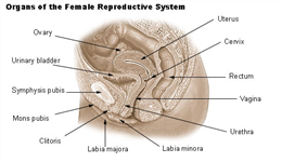
The vulva is the external genitalia in the female reproductive system.
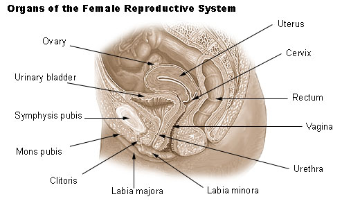
Broadly the anatomical structures of vulva include:
- The lips of the vulvar opening – Labia major and labia minora
- The swelling at the upper part of the vulvar opening or the mons pubis
- The seat for nerve endings and sexual stimulation in females or the clitoris
- The sac like areas called the bulb of the vestibule and the vestibule of the vagina
- The greater and lesser vestibular glands, Bartholin gland
- And the vaginal orifice
- The urethral opening
Anatomical features of the vulva
- Vestibular area – This lies between the hymen and Hart's line. There are linings with non-keratinized squamous epithelium. Within the vestibule are major vestibular (Bartholin's) glands, the minor vestibular glands, the periurethral (Skene's) glands, and the urethra.
- Labia majora – These begin from the walls of the vulva. They are composed of folds of adipose (fatty) and fibrous tissue. These join at the front of the vulva to form the mons pubis. Behind the vulva they terminate 3-4 cm in front of the anus where they are united by the posterior commissure or fourchette. The cleft between the ''labia majora'' is called the pudendal cleft, or ''cleft of Venus'', and it contains and protects the other, more delicate structures of the vulva. These are lined by skin on the outside and basal layer of cells (stratum malpighii), a thin granular layer on the insides.
- Labia minora – These are two folds of connective tissue which contain little or no adipose tissue. In the front the labia minora divide into two parts. One part passes over the clitoris to form the prepuce. The other joins beneath the clitoris and forms the frenulum. Behind they blend with the inner surfaces of the labia majora. The skin and mucosa of the labia minora are rich in sebaceous glands. These glands secrete fluids to maintain lubrication.
- Hymen – The introitus is sometimes partly covered by a membrane called the hymen. The hymen will rupture during the first episode of vigorous sex, and the blood produced by this rupture is seen as a sign of virginity. However, the hymen may also rupture spontaneously during exercise or be stretched by normal activities such as use of tampons
- The urethral opening within the vulva – The urethra opens into the vestibule anterior to the vaginal introitus. The cells that line the urethra are called transitional epithelium with stratified squamous epithelium at the orifice or opening. The muscles of the tube like urethra are a longitudinal inner layer and a circular outer layer of smooth muscles.
- Skene's ducts – These are at the floor of the end of the urethra and open just within or external to the opening. These are 0.5 to 1.5 cm long
- Bartholin's gland – This is similar to the bulbourethral glands in the male. The gland ends in a tube that is lined by transitional epithelium.
- Sebaceous glands – These glands lie along the inner parts of the labia minora. These look like raised spots. There are no hair follicles around these glands.
- Other apocrine glands – These are scent glands that are identical to those of the axillae, breast and perianal regions.
- Other eccrine glands – These are sweat glands that are primarily involved in heat regulation. They function before puberty. They are lined by a layer of epithelial cells that contain an eosinophilic cytoplasm.
- The mons pubis – This is the fatty cushion resting over the front surface of the pubic bone. This area is covered with pubic hair after puberty.
- The clitoris – This is a small cylindrical erectile tissue similar to the penis in males. It has a rich supply of blood and nerves. Even though the clitoris is much smaller than the penis, it has twice the amount of nerve endings than the penis.
- The perineum – This is the tissue between the vagina and the anus. During childbirth it may stretch or tear.
The appearance of the vulva and the size of the various parts varies a great deal from one female to another. The left and right sides of the external genitalia may also differ.
Sources
- www.asccp.org/…/Default.aspx
- http://www.nva.org/vulvarAnatomy.html
- www.midirs.org/…/The%20Vulva.pdf
- www.urmc.rochester.edu/…/wk11d-AnatomyoftheVagina.pdfhttp://www.pinkrobin.co.uk/shop/Anatomy.pdf
Further Reading
- All Vulva Content
- What is the Vulva?
- Vulva Development
- Vulva Sexual Arousal
- Vulva Fluids and Odor
More…
Last Updated: Jun 5, 2019

Written by
Dr. Ananya Mandal
Dr. Ananya Mandal is a doctor by profession, lecturer by vocation and a medical writer by passion. She specialized in Clinical Pharmacology after her bachelor's (MBBS). For her, health communication is not just writing complicated reviews for professionals but making medical knowledge understandable and available to the general public as well.
Source: Read Full Article