What is Somitogenesis?

How are the body’s segments formed?
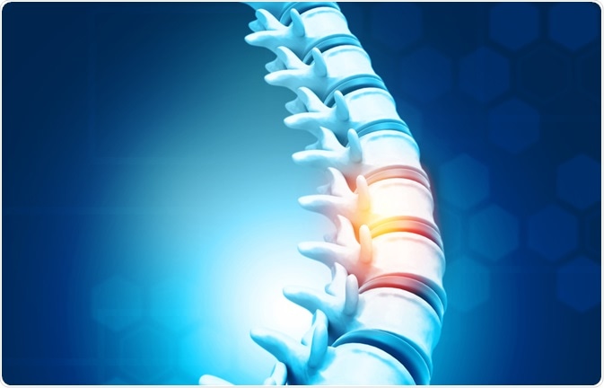
The body of a vertebrate organism consists of segmented axes, which is most distinct in the skeleton. The process of somitogenesis is key to the development of these segments, or somites, which then develop into vertebrae and skeletal muscle.
Image Credit: Explode/Shutterstock.com
Generally, the process involves the creation of the presomitic mesoderm, formation of the somitomeres, the formation of the epithelial somites followed by their differentiation into dermatome, myotome and sclerotome, and then their morphogenesis according to their position in the developing embryo.
How does the presomitic mesoderm form?
Gastrulation, where embryonic cells migrate through a blastopore and subsequently organize to form the three germ layers of the embryo, is when the process of somitogenesis begins. During gastrulation, cells undergo an epithelial to mesenchymal transition, regulated by the transcription factor Snail.
Studies using mice and chick embryos have shown that certain cells become specified and are then maintained within a domain of the blastopore, including progenitors which will form the presomitic mesoderm.
These cells then proliferate and migrate away from the blastopore and position themselves in the posterior region of the presomitic mesoderm. This migration serves to replace cells as somites bud off from the anterior region of the presomitic mesoderm.
What is the segmentation clock?
Within the presomitic mesoderm, there is a wave of gene expression patterns that are driven by synchronicity across the cells, and this matches somite formation. This wave begins at the posterior end, and when the wave reaches the anterior end a somite pair buds off. This subsequently becomes the signal for a new wave to begin at the posterior end, thus creating a clock-like pattern.
The determination front, where cells become segments
Within the presomitic mesoderm, there is a gradient of Fgf8 and β-catenin expression that runs in the posterior to anterior direction, which is in contrast to the activity of retinoic acid that runs in the anterior to posterior direction.
The point where these gradients meet has been termed the “determination front” and is where the presomitic mesoderm begins to segment. It is believed that the size of these segments is determined by the number of cells that pass through the determination front during one cycle of the segmentation clock.
How are boundaries between somites formed?
Boundaries need to be formed within the somites for proper segmentation. This occurs at the anterior end of the presomitic mesoderm and involves a cascade of gene expression driven by Notch activity during anterior migration.
Notch activates the Mesp2 gene in the somite anterior to the determination front, but this is inhibited by FGF in the cells posterior to the determination front. Mesp2 expression is then refined to make the somite polar.
Mesp2 then causes more adhesion proteins to be expressed, which leads to the cells at the segment boundary to be epithelialized and form the boundary between somites. This is where the epithelial somites are formed.
Becoming something new; how do somites differentiate?
The maturation and differentiation of somites into the dermatome, myotome, and sclerotome depend on the signals from the surrounding cells.
– Cells in the dorsal part of the somite are close to the ectoderm and the neural tube, and the expression of Wnt1/3a from the neural tube and Wnt8c from the ectoderm induces the expression of Pax3 and Pax7 in the somite. This means that these cells remain epithelial, and they form the dermomyotome.
Cells away from the tip of the dermomyotome are then influenced by the expression of NTF3 by the neural tube, which drives this section of the dermomyotome to become the dermatome. The dermatome will later form the skin.
Cells at the tip of the dermomyotome, on the other hand, become the myotome, which will later form the muscles in the body and back, as well as skin in the back.
– Cells in the ventral part of the somite become de-epithelialized, which is driven by the low levels of sonic gene (Ssh) expression in this part of the somite. This then becomes the sclerotome, which will later form the vertebrae and tendons. The expression of Hox genes dictates the differences in the shape of the vertebrae along the anterior-posterior axis.
What happens when somitogenesis goes wrong?
Somitogenesis is a crucial process during human embryonic development, and certain skeletal and muscular deformities have been linked to defects during somitogenesis.
While the origin of most of these defects remains unclear, mutations within LFNG, MESP2, HES7 and delta-like 3 have been linked to some of these defects; these genes are known to be important during early presomitic mesoderm formation.
Sources
- Maroto, M. et al. (2012) Somitogenesis. Development https://dev.biologists.org/content/139/14/2453
- Gossler, A. and de Angelis, M. H. (1997) Somitogenesis. Chapter 6, Current Topics in Developmental Biology vol. 38 https://www.sciencedirect.com/science/article/pii/S0070215308602483
Further Reading
- All Embryonic Development Content
- What is Gastrulation?
- The Stages of Early Embryonic Development
- An Overview of Morphogens and their Signaling Pathway
Last Updated: Mar 26, 2020

Written by
Dr. Maho Yokoyama
Dr. Maho Yokoyama is a researcher and science writer. She was awarded her Ph.D. from the University of Bath, UK, following a thesis in the field of Microbiology, where she applied functional genomics toStaphylococcus aureus . During her doctoral studies, Maho collaborated with other academics on several papers and even published some of her own work in peer-reviewed scientific journals. She also presented her work at academic conferences around the world.
Source: Read Full Article
