Serial Block Face Scanning Electron Microscopy Applications
Serial block-face scanning electron microscopy (SBEM) is a type of scanning electron microscope that has an ultramicrotome mounted inside its vacuum chamber.
 Credit: Life science/Shutterstock.com
Conventional microscopy cannot resolve some cellular structures, and does not capture three-dimensional features of the sample. The connectivity of local networks of neurons is an example of information that could be lost in a biological tissue under an electron microscope or a light microscope.
Serial block-face scanning electron microscopy (SBEM) was invented in 2004 by Winfried Denk of the Max Planck Institute in Heidelberg. It was originally developed to analyze connectivity of axons in the brain, but scientists have since found wide applicability for many types of biological samples.
In SBEM, the sample is fixed and stained and the surface is imaged by detection of back-scattered electrons. Then a thin section of about 30 nm is cut from the face of the block and it is imaged again.
Neuroscience
SBEM is an important application used in neuroscience for understanding the neural networks within the brain. The following new approaches have been implemented:
- In-vivo imaging: Various aspects of cell structure and function have been studied using SBEM, including stem cell differentiation, transformation, axons, functional and structural neuroplasticity, networks, healing, and growth have been carried out using SBEM.
- Brain mitochondria: SBEM has been used to study mitochondrial structures in mammalian brain tissues, including shape, number of mitochondria, volume, size, and location. The high resolution of the technique enables identification of all of the structures within the mitochondria, as well as structural defects.
Human chromosomes
The structure of human chromosomes within the 30 nm range is poorly understood. Techniques such as transmission electron microscopy (TEM) and scanning electron microscopy (SEM) are not able to probe the internal structure of a chromosome.
However, SBEM has been used to visualize the chromosomes in mitotic cells. The results showed clear signs of internal structure. Images were detailed enough to reveal internal pores of the chromosome. Resolution was limited to about 11 nm.
Cardiac muscular structures
SBEM studies of cardiac muscles reveals structural details of the tissue and images of cardiac myocytes present in the myocardium, as well as mapping cellular structures and interconnections between transverse tubules and sarcoplasmic reticulum. It also gives quantitative information including volumes occupied by the various structures.
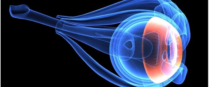
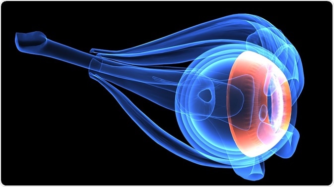
Ophthalmology
Degenerative conditions of the retina often present with subtle phenotypic changes before the disease is fully manifested. To observe those events, high resolution of large areas of the retina is needed.
SBEM can be used to image large volumes of retinal tissue at the necessary resolution. In a study by Mustafi et al, researchers identified phenotypic changes preceding onset of disease in mouse models of various human retinal dystrophies.
Material science
SBEM is widely used in imaging biological structures due to their complexity. However, it also has utility for solving problems in material science, such as optimizing microfiltration membranes used in recycling of wastewater and analyzing metallic coatings.
SBEM can also be used to analyze 3D crystal structures of materials and combined with energy dispersive X-ray, and to engineer nanoscale 3D chemical textures.
Sources:
- Developing 3D SEM in a broad biological context
- Serial block face scanning electron microscopy for the study of cardiac muscle ultrastructure at nanoscale resolutions
- 3D imaging by serial block face scanning electron microscopy for materials science using ultramicrotomy
- Serial block face scanning electron microscopy: a method to study retinal degenerative phenotypes
Further Reading
- All Electron Microscopy Content
- Electron Microscopy: An Overview
- Applications of Electron Microscopy
- Advantages and Disadvantages of Electron Microscopy
- What is Transmission Electron Microscopy?
Last Updated: Feb 26, 2019

Written by
Dr. Catherine Shaffer
Catherine Shaffer is a freelance science and health writer from Michigan. She has written for a wide variety of trade and consumer publications on life sciences topics, particularly in the area of drug discovery and development. She holds a Ph.D. in Biological Chemistry and began her career as a laboratory researcher before transitioning to science writing. She also writes and publishes fiction, and in her free time enjoys yoga, biking, and taking care of her pets.
Source: Read Full Article